Cracking the code on brittle materials

A close-up image shows an array of white dots. The movement of these dots represents the change in surface slope of cracking glass.

Blink and you’ll miss it: an array of flickering dots, each one mere microns wide, represents the lightning-fast expansion of a crack in glass as a barely perceptible shimmer. 

Using an instrument and approach typically used in medicine and astronomy, a team of Hopkins Extreme Materials Institute (HEMI) researchers has tackled a longstanding challenge in fracture mechanics: understanding and quantifying the behavior of fast cracks in brittle materials like glasses and ceramics.

Led by postdoctoral fellows Liuchi Li and Velat Kiliç, the team used Shack-Hartmann wavefront sensing (SHWFS)—a decades-old method for detecting ophthalmic problems and correcting distortion in telescope images—to map changes in shape and movement that happen in material surrounding rapidly spreading cracks with much higher precision and on much faster timescales than ever before. Their approach appears in Mechanics of Materials.

“Can we make glass that won’t crack? How can we make materials last longer? Once cracking begins, how can we prevent a structure’s total failure? Our unconventional use of this old method enables scientists to tackle these important questions,” said Li.

Making stronger, more resilient glass requires a better understanding of stress intensity factors (SIFs) in highly brittle materials. A measure of stress around or near a crack or flaw in a material, SIFs allow scientists to quantify and predict how cracks evolve. In brittle materials, deformations are very small and the speed at which the ends or edges of cracks as they spread are high, making SIFs difficult to determine using direct measurements.

While theories and simulations allow scientists to predict SIFs, high-resolution experimental data has so far been scarce. One established method, digital image correlation (DIC), can sometimes miss small changes. Another, digital gradient sensing (DGS), is better at tracking those small changes but does not capture as much detail.

Mark Foster, co-author and associate professor of electrical and computer engineering, suggested the team explore SHWFS as a way to map both overall changes and small details in brittle materials under stress.

“Shack-Hartmann wavefront sensing seemed like the ideal solution due to its ability to directly measure surface profiles with high spatial resolution. One thing that is particularly unique here is that we are implementing wavefront sensing at extraordinary frame rates to capture the SIF information,” said Foster.

Both DGS and SHWFS use light to measure surface deformation. However, the Shack-Hartmann sensor used by Li and his colleagues has a spatial resolution of about 10 microns—a hundred times better than the resolution typical of conventional DGS. (For reference, a single human hair has a diameter of around 50 to 100 microns.) This allows the team to get remarkable data about behavior near the crack tip, even though it is moving at hundreds of meters per second.

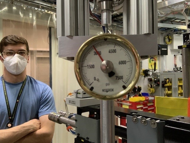
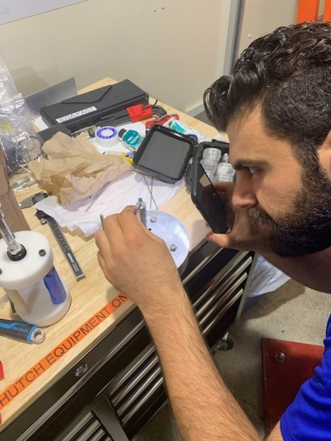
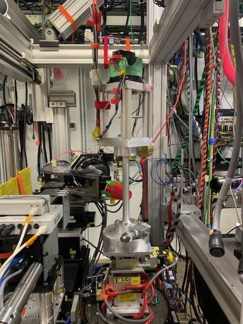
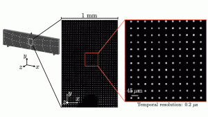
The team’s SHWFS setup, pictured below, uses a series of lights, lenses, and cameras to transform beams of light into an array of dots. As a crack spreads through a sample, the dots in the array change position slightly, representing changes in the sample’s surface slope. The team compared their results to full-scale 3D finite element simulations to verify their method and experimental measurements.

An experiment setup is shown. Figure A) Diagram: a collimator shines light onto a sample, which is reflected and magnified using a series of lenses and a microlens array. The light reaches an image plane, where a relay and camera collect visual information. Figure B) An image of the setup described in figure A is shown. The equipment is arranged on an optical table with screwdrivers and wires scattered around. Figure C) The data collected by this setup is represented by an array of white dots. The enlarged image shows a 1mm by 1.6mm sample.

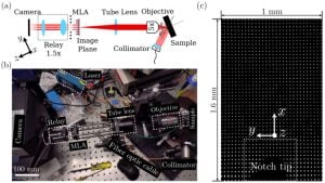
The team’s SHWFS setup (figures a and b) focuses beams of light through a microlens array to provide an average slope of the sample’s surface over a given area. These slope measurements are represented by an array of dots (figure c), which is captured as a series of images by a camera. As the sample cracks, the dots shift slightly to reflect deformations in its surface. A computer program developed by Li computes the shifts in each dot’s position from one snapshot to the next.

“Our method’s unprecedented spatial resolution allows us to probe fundamental questions in the fracture mechanics of brittle materials, opening the door for predictive dynamic fracture theories for engineering applications,” said Li.

The researchers say that their method could not only inform guidelines for civil engineers but also help in the development of novel materials for consumer electronics and in the defense and aerospace sectors.

“An important next step is to integrate SHWFS with complementary techniques,” says Todd Hufnagel, co-author and professor of materials science and engineering. “For example, x-ray phase-contrast imaging (XPCI) can reveal the precise shape and orientation of the propagating crack tip. The combination of the two techniques will give us unprecedented insight into the nature of dynamic crack propagation in brittle materials.”

Team members included researchers from the departments of Electrical and Computer Engineering, Materials Science and Engineering, and Mechanical Engineering at the Whiting School. In addition to Li, Foster, and Hufnagel, authors include KT Ramesh, Alonzo G. Decker Jr. Professor of Science and Engineering, and HEMI postdoctoral fellows Milad Alemohammad and Lei Yang.

ACKNOWLEDGEMENT STATEMENT:

This project was sponsored in part by the Department of Defense, Defense Threat Reduction Agency under the Materials Science in Extreme Environments University Research Alliance, HDTRA1-20-2-0001. It was also supported in part by the Corning Research and Development Corporation.

Rethinking the building blocks of soil mechanics

Understanding how granular materials and rocks react under stress is crucial to projects ranging from building stable roads and bridges to formulating pills and cosmetics. Soil and rock mechanics are also integral to academic fields such as civil engineering, materials science, geology, and seismology.

When granular materials experience stress, they can start to behave in a fluid-like manner. This behavior is known as plastic flow, and plasticity theories attempt to explain this and related phenomena. Although plasticity theories have been developed and used for decades, there have been no direct stress or strain measurements to validate them, in part because of the significant challenges in directly measuring stress deep within a deforming material.

Using advanced imaging techniques and a unique experimental approach, researchers at the Hopkins Extreme Materials Institute have collected data that is among the first of its kind. Ryan Hurley, assistant professor of mechanical engineering in the Whiting School of Engineering, leads a research group studying the mechanical behavior and failure mechanisms of various geomaterials. Hurley and his collaborators (including mechanical engineering doctoral students Brett Kuwik and Kwangmin Lee and former HEMI postdoctoral fellow Ghassan Shahin) have recently had their findings published in Proceedings of the National Academy of Sciences.

During this study, the team performed triaxial compression tests on synthetic quartz sands, with x-ray imaging and diffraction offering some of the first known in-situ grain stress and local strain measurements.

Using a specialized triaxial compression instrument developed in Hurley’s lab, the researchers placed quartz sand samples under confining pressures between 20 and 35 MPa—2900 to 5000 pounds per square inch (PSI)—and analyzed them using synchrotron x-ray tomography and 3D x-ray diffraction at the Advanced Photon Source at Argonne National Laboratory.

“Results from this study provide the first direct confirmation of some of the fundamental postulates we use when studying the plastic deformation of sands,” said Hurley. “On the other hand, our results contradict prior assumptions such as the existence of ‘dead zones’ outside of regions of localized straining, indicating that some fundamental ideas about plastic flow in granular materials may need to be revisited.”

The team says these findings provide an opportunity for scientists to begin evaluating certain assumptions about how granular materials such as sand respond to stress, re-evaluate old ideas, and validate and expand upon existing models in soil and rock mechanics.


3D x-ray images of sand before and after axial straining, along with renderings of the sample. Sand particles are colored by their motion (instantaneous rotations) and stress. See Hurley’s paper in this edition of PNAS for additional details.

News flash: Study decodes secrets of impacts in space

Impacts by debris and meteoroids pose a significant threat to satellites, space probes, and hypersonic craft. Such high-velocity impacts create a brief, intense burst of light, known as an impact flash. These flashes contain information about both the target and the object that caused the impact. For example, scientists have studied the impact of meteoroids on the moon by monitoring and analyzing impact flashes on its dark side.

A team of Johns Hopkins Whiting School of Engineering researchers led by mechanical engineering doctoral student Gary Simpson, Professor of Science and Engineering K.T. Ramesh, and colleagues have discovered that impact flashes are created by the fragmentation of an ultra-fast jet of material ejected from the colliding bodies. Their results appear in PNAS Nexus.

Working in the Hopkins Extreme Materials Institute’s HyFIRE lab, the team shot stainless steel spheres into an aluminum alloy plate at a speed of three kilometers per second—about 6,700 miles per hour, or more than nine times the speed of sound. The resulting impact flashes were photographed using ultra-high-speed cameras and high-speed spectroscopy, which measures the color and brightness of the light.

Immediately after impact, a luminous disc is seen expanding around the impacting sphere. Only a few millionths of a second later, the disc takes on an almost floral shape, as fragments ejected from the impact crater form an ejecta cone, with petal-like projections at the outer edge.

“We found that minuscule, condensed fragments from the jet interact with the atmosphere to create an extremely bright radiating cloud of vapor, which expands at a speed of over ten kilometers per second, or more than 22,000 miles per hour,” said Simpson. “The material making up the target and the size of the jetted particles can be inferred from the flash.”

Other authors of the study include Justin Moreno, HEMI associate staff engineer, and Matthew Shaeffer, HEMI senior staff engineer. Both are faculty members in applied biomedical engineering at the Whiting School’s Engineering for Professionals program.

HEMI Fellows awarded best papers at MS&T conference

Lori Graham-Brady, HEMI associate director and professor in the Department of Civil and Systems Engineering and KT Ramesh, the Alonzo G. Decker Professor of Science and Engineering and director of HEMI, were awarded best papers by the Journal of the American Ceramic Society. On October 11, 2022, their winning papers were presented at a special awards symposium at the Materials Science and Technology Technical Meeting and Exhibition in Pittsburgh, Pennsylvania.   

Graham-Brady’s paper is titled, “Fragmentation and Granular Transition of Ceramics for High Rate Loading,” and included co-authors Amartya Bhattacharjee and Ryan Hurley of Johns Hopkins University. 

“Models for the Behavior of Boron Carbide in Extreme Dynamic Environments,” is the title of K.T. Ramesh’s winning paper. Co-authors included: Lori Graham-Brady, Ryan Hurley, Mark Robbins, Amartya Bhattacharjee, Qinglei Zeng, Weixin Li, and Nilanjan Mitra from Johns Hopkins University; William Goddard, California Institute of Technology; Andrew Tonge, DEVCOM Army Research Laboratory; Joel Clemmer, Sandia National Laboratories; and Qi An, University of Nevada, Reno. 

Both papers were the result of research conducted in the Center for Materials in Extreme Dynamic Environments(CMEDE), a center within the Hopkins Extreme Materials Institute. Funded by the DEVCOM Army Research Laboratory, CMEDE research has developed a materials-by-design process for protection materials which have military armor applications.    

HEMI Fellow Sarah Hörst Featured in New York Times for Exoplanetary Research

Sarah Hörst, HEMI Fellow and associate professor in the Department of Earth and Planetary Sciences, was recently featured in a New York Times article titled, “You Don’t Need a Spaceship to Grow ‘Weird Little’ Martian Radishes”.

The piece showcases Hörst’s research on Saturn’s moon, Titan. It also speaks to the work done in her lab regarding hypothetical exoplanets, which is helping to figuring out which potential exoplanets produce smog. This information can help scientists point telescopes at orbs they can actually observe.

Sarah Hörst of Johns Hopkins University with her lab’s planetary atmospheric simulation chamber.Credit…Justin Tsucalas

In 2018, Hörst mentored a student in the HEMI/MICA Extreme Arts Summer Project who used their art to help explain the intricacies of Titan’s atmosphere. Since that time, Hörst has grown her role and  is currently leading the program’s development.

Sarah Hörst (JHU) and Amy Wetsch (MICA) at the 2018 opening of Wetsch’s show “Lateral Distance.” The show featured pieces meant to artistically visualize and simulate Titan’s atmosphere. (image: Will Kirk, Homewood Photography)

HEMI Graduate Student Suhas Eswarappa Prameela Featured in Science Working Life

Suhas Eswarappa Prameela, a graduate student in HEMI, has been featured in Science Magazine’s Working Life section with an essay about finding his voice on Twitter.

His essay describes how he found value in sharing his experiences on the science side of Twitter. In the beginning, he only tweeted about his research results. One day, out of frustration, he tweeted about the “importance of properly naming research files.” Prameela was retweeted by a high-profile academic account and went viral overnight.

Prameela initially had apprehensions about sharing his experiences online. He said, “Who was I to offer advice? I’m just a PhD student doing my best; I didn’t know whether I was doing anything right myself!” However, after falling into his new-found Twitter fame, he began tweeting regularly about his personal experiences as a PhD student: from navigating authorship conflicts to giving presentations and more.

As a result of his efforts, other students on Twitter have reached out to him, thanking him for his tweets and asking questions. Prameela says that although sometimes he feels “uncomfortable and vulnerable sharing my experiences on a massive global platform, I’ve found it is well worth it. I’ve learned that we are all stronger if we authentically share and connect with one another.”

Science Magazine’s Working Life section features articles that discuss the professional side of science and research, with topics ranging from mentorship to mental health struggles and more. Click here to read the full text of Prameela’s essay.

You can follow Prameela on Twitter (@suhas_prameela) here.

Simulations offer new approach for designing high strength metals under high rate loading

Jaafar El-Awady, HEMI Fellow and associate professor of mechanical engineering, has co-authored research that reveals more clues about the microscopic mechanisms that govern the strength of metals.

In the paper recently published in Nature Communications, titled “Strain rate dependency of dislocation plasticity,” El-Awady and team present a new understanding of the effects of strain rate and dislocation density on the deformation behavior of aluminum and cooper.

Metals like aluminum and cooper are commonly used engineering materials, thanks in part to their strength and damage tolerance. Why, then, do metals sometimes deform or break unexpectedly due to stress? Understanding why metal components fail will inform the design of stronger metals, improving their performance in a broad range of applications.

In materials research, the flow strength refers to a material’s ability to resist deformation under an applied load. Metal properties are loading rate sensitive, meaning they react differently across various rates of loading. Dislocations, or line defects, in metals also move and localize under loading, which is a precursor to crack initiation and failure. Thus, researchers hypothesize that dislocation density, or the number of dislocations per unit area in a material, may affect the loading rate sensitivity of the material. However, the relationship between the two mechanisms is still not well understood.

To solve this problem, the team performed a large set of three-dimensional discrete dislocation plasticity simulations to predict the metal yield strength at different initial dislocation densities and strain rates. The simulation results showed that the material response can be divided into two regimes. In one regime, dislocation-dislocation interactions control the material flow strength; in the other, strain rate induced hardening dominates. Additionally, the transition between both regimes is mainly controlled by the dislocation density in the material.

Based on their observations, the team was able to develop a unified law that correlates the material flow strength with the loading strain rate and the initial dislocation density in the material. This unified law is shown to be in excellent agreement with a large set of experimental results from previous studies.

The study provides a fundamental understanding of the co-interaction of different mechanisms in materials and how they influence the material strength, said El-Awady. These findings could help researchers design metals better suited for high strain rate loading applications, by tailoring the initial dislocation density in the material to achieve a desired strength.

In addition to El-Awady, the research team includes first author Haidong Fan, a former Johns Hopkins postdoc and current professor at Sichuan University, Qingyuan Wang, Dierk Raabe, and Michael Zaiser.

Excerpted from the Department of Mechanical Engineering >>

HEMI Fellow Sung Hoon Kang Highlights Applications for Microactuators in Science Robotics Article

Sung Hoon Kang, HEMI Fellow, assistant professor in the Department of Mechanical Engineering, and associate researcher at the Institute for NanoBioTechnology, has written a focus article about the fabrication of and potential applications for an electrically-controlled shape-memory microactuator that operates in a solution matching the ion concentration of the human body. The article was published in Science Robotics and includes contributions by Kang’s graduate students Mostafa Omar and Bohan Sun.

Titled “Good reactions for low-power shape-memory microactuators,” the article details the mechanism of the microactuator and its potential versatile applications. The microactuator has two configurations: bent and flat. When the bent microactuator is exposed to an electrochemical oxidation reaction, an oxide layer grows on its surface, expanding and flattening the device into its flat configuration. The oxide layer stays after the reaction, which allows the device to maintain its shape without applying voltage. As such, it is especially useful for robots operating with limited power supplies.

The article showcases the versatility of microactuators in extreme environments. For example, in medicine, applications can include: controlled drug delivery, temporary clamps in place of stitches, and fast-response devices against blood clots in arteries. Other applications include use as a microvalve to control flow in microfluidics and reconfigurable optical metamaterials/metaservices. Its potential for application can be further increased by improving energy efficiency, speed, actuation range, and durability.

Read the focus article here.


HEMI Fellow Jaafar El-Awady Identifies New Method to Predict Cracks in Metal Before They Are Visible

Congratulations to Jaafar El-Awady, HEMI Fellow and associate professor in the Department of Mechanical Engineering, for discovering a new method to predict cracks in metal before they appear. His results were published in Science.

His paper, titled “The heterogeneity of persistent slip band nucleation and evolution in metals at the micrometer scale,” identifies a new method to test metals and predict vulnerabilities before visible cracks are formed. This has implications in understanding and predicting the life of a metal mechanism, especially to help prevent fatigue failure and to avoid discarding parts that may still be useful.

“We’re able now to have a more fundamental understanding about what leads up to cracks,” El-Awady said. “The practical implication is that it will allow us to understand and predict when or how the material is going to fail.”

Read El-Awady’s paper here.